.png) 0361-2800800
0361-2800800 (+91) 9864103333


Sonography (Ultrasound)
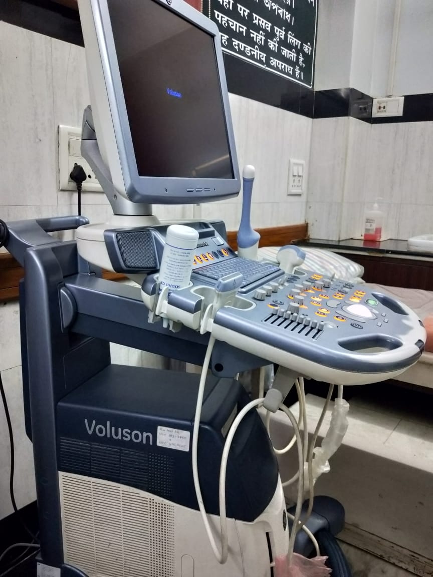
Sonography is one of the basic and essential diagnostic services. Our ultrasound department is equipped with a very high-end ultrasound machine of the Voluson series. Apart from routine ultrasound, the machine is equipped to do Colour Doppler, Power Doppler, 3D, and 4D ultrasound. All basic and advanced ultrasound which are required for mother and child care is performed at IHR on daily basis.
Advanced ultrasound methods required for assessment of infertile couple such as 3D TVS, 3D HISTOGRAM to assess uterine receptivity, scrotal ultrasound are also done every day at IHR.
Pregnancy detection, ectopic pregnancy identification, the viability of the pregnancy, aneuploidy scan, anomaly scans, growth assessment, Neuro-sonogram of a new born are also done here.
Preparation
-
For abdomen sonography, 4 hours of fasting is required.
-
For pelvic sonography (Trans-abdominal sonography: TAS) full bladder is required which can be achieved by not passing urine for at least 1 hour before the scan and drinking about 1 litre of water during this time.
-
For transvaginal sonography (TVS), no special preparation is required.
-
For pregnancy ultrasound NO special preparations are required, i.e. no empty stomach or no full bladder is required if you are pregnant.
-
Follicular study is done using TVS, so no special preparations are required.
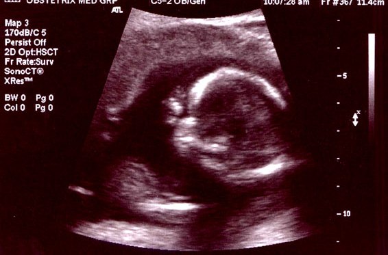
Routine Pregnancy Scan
Ultrasound is a very useful technique during pregnancy to rule out any congenital abnormality in the growing fetus inside the uterus. Not just the health of the baby, but also the health of the to-be-mother can be determined with the help of ultrasounds. It helps in measuring the growth of the baby in terms of her weight and height. Gynecologists recommend regular ultrasounds to check the levels of amniotic fluid, to rule out multiple pregnancies or ectopic pregnancy, to estimate delivery date, and to measure cervical length as well.
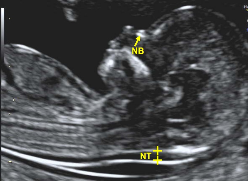
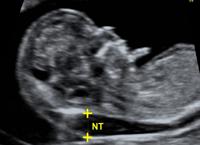
Aneuploidy/ Nuchal Translucency(NT) scan
Nuchal translucency scan is done around the 11th to 14th week of pregnancy. The scan aims to measure the size of the nuchal fold at the back of the baby’s neck to rule out the possibility or risk of any chromosomal abnormalities. With the help of a nuchal translucency scan, your health professional will rule out the risk of abnormalities like trisomy 21 (Down Syndrome), trisomy 18 (Edwards Syndrome), or trisomy 13 (Patau syndrome).
Fetal Echocardiography
Fetal Echocardiography is a test similar to ultrasound. It allows the doctor to see the structure and function of the baby’s heart in detail. It also enables the doctor to see blood flow through the fetal heart and cardiac rhythm.
Click Here to Know More
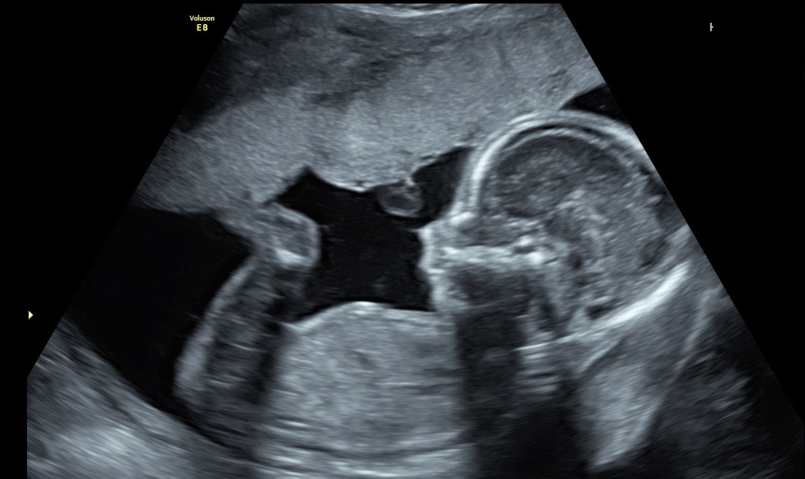
Anomaly Scan
Anomaly Scan or mid-pregnancy scan is an ultrasound scan done between the 18th and 21st week of pregnancy to take a closer look at the baby and the womb (uterus) and to have an idea where the placenta is lying. This scan aims to look for any major physical abnormalities in the growing baby. Although it can’t pick up every problem, it gives an idea about the baby’s bones, heart, brain, spinal cord, face, kidneys, and abdomen and allows the identification of the following conditions (some of which are very rare):
| Brain/Skull |
|
| Face |
|
| Chest |
|
| Abdomen |
|
| Spine |
|
| Extremities |
|
| Brain/Skull |
|
| Face |
|
| Neck |
|
| Chest |
|
| Heart |
|
| Abdomen |
|
| Genitourinary |
|
| Spine |
|
| Extremities |
|
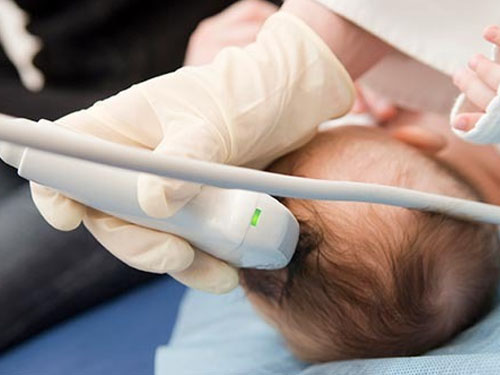
Paediatric Scan
A pediatric ultrasound is an examination of the child’s abdomen with the help of an ultrasound machine. The procedure involves an ultrasound gel and a small probe (transducer) that uses sound waves to produce an image. It is a non-invasive medical examination that helps our experts diagnose and treats abdominal pain and investigate other useful information about other organs such as the liver and kidneys. This helps determine the causes of vomiting in young infants, detect the cause and the presence of an apparent enlarged abdominal organ, guide other procedures including biopsies and identify the location of abnormal fluid in the abdomen.
Sonography (Ultrasound) Team

Interventional Radiology
IHR offers a wide range of Ultrasound (USG) guided procedures under one roof for the convenience of the patients. All the procedures are conducted by the experts, using the latest and advanced equipment. They are daycare procedures and the patient is fit to go back home the same day.